|
Pelco Graphene products are available on a variety of substrates including lacey carbon, 300 mesh Cu grid, 2000 mesh Cu grid, holey silicon nitride, and ultra-flat SiO2. The Graphene is available in different thicknesses: single layer, 2-layers, 3-5, and 6-8 layers. Graphene sheets with thicker layers are available by special order.
Graphene on Lacey Carbon
The PELCO® Graphene TEM Support Films are suspended on a lacey carbon film on a 300mesh grid (#01895). The Graphene films we offer have either single, 2, 3-5, or 6-8 layer Graphene sheets and cover the entire TEM grid. The usable area is around 75% due to some unavoidable folds and wrinkles in the Graphene sheets. Graphene, with its unique properties, offers a support film layer that is more conductive and also much thinner than the average carbon support film. Although it is a crystalline support film, its contribution to signal formation is relatively low. This makes the single and 2-layer Graphene support film ideal for high resolution imaging, imaging of nanoparticles and imaging of weak contrast materials/interfaces.
Graphene Specifications
- The sheet resistance for a single layer of Graphene Film is 600Ω/sq.
- The thickness for a single layer of Graphene is approximately 0.35nm, Transparency is in the order of 96.4%.
- The thickness for 2 layers of Graphene is approximately 0.7nm, Transparency is in the order of 92.7%.
- The thickness for 3-5 layers of Graphene is between 1.0 - 1.7nm, Transparency ranges from 90.4 - 85.8%.
- The thickness for the 6-8 layers of Graphene is between 2.1 - 2.8nm, Transparency ranges from 83.2 - 78.5%.
The PELCO® Graphene has an in-plane modulus of 0.9TPa, compared to 1.0 TPa for Graphene produced by the Scotch Tape™ method.
 |
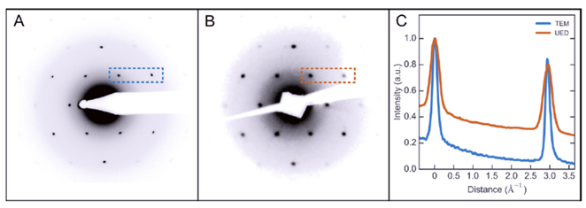
Comparison of the diffraction pattern of Graphene on copper mesh coated with a lacey carbon film measured with (a) a transmission electron microscope (TEM) at 80 kV and (b) the ultrafast electron diffraction (UED) setup at 6 kV. The line profiles in (c) correspond to the highlighted regions in the diffraction patterns in (a) and (b). These data have been normalized to the intensity of the first order peak. The widths and locations of neighbouring Bragg peaks were used to estimate the transverse coherence length of the UED setup. Ref: Badali, et. al. Struct. Dyn. 3, 034302 (2016). |
|